Microsporidial Keratoconjunctivitis
Summary
Microsporidial keratoconjunctivitis (MKC), once a rare opportunistic infection centred on AIDS patients, is now increasing worldwide as a keratoconjunctivitis of immunocompetent people — especially outdoor-sport athletes. Its occurrence has been shown to be strongly governed by hot, rainy and heavy-rain conditions, and the disease is increasingly viewed as a "climate-sensitive infection" to be monitored under climate change.
- Disease Review (Part I): the pathogen is a spore-forming microbe related to fungi, and the main cause of ocular infection is Vittaforma corneae, among others. It is transmitted via soil and water and is culture-negative and poorly responsive to standard treatment, so it is easily misdiagnosed or overlooked (e.g. as adenoviral keratoconjunctivitis).
- Evidence from Japan & Domestic Projections (Part II): in two outbreaks in Nagasaki (2022 and 2023; 16 healthy athletes in total), the same V. corneae was detected molecularly in patients' corneas and in playing-field soil — direct evidence linking the environmental reservoir (soil and water) to human infection. Based on JMA data, intensifying heat and heavy rain are expected to expand suitable areas within Japan from Kyushu toward Kanto, Tohoku and Hokkaido.
- Outbreak projections worldwide (Part III): using the same meteorological threshold and IPCC AR6 data to gauge suitable areas, the monsoon belts of South and Southeast Asia and tropical Oceania are already high-risk, and climate change is expected to spread outbreaks from the tropics and subtropics into the temperate zone.
I-1. Introduction
In the differential diagnosis of keratoconjunctivitis, viral (especially adenoviral) and bacterial causes are common, whereas microsporidial infection is an easily overlooked cause that has attracted attention in recent years. Once regarded as a rare opportunistic infection, the disease has been reported with markedly increasing frequency as a cause of keratoconjunctivitis in immunocompetent people, in step with advances in diagnostic technology and greater clinical awareness. In particular, onset has been noted to be associated with the rainy season and heavy rainfall, raising concern that the increases in rainfall, flooding and hot, humid conditions accompanying global warming may affect the frequency and geographical distribution of the disease. Part I organizes the pathogen biology, epidemiology, clinical features, diagnosis and treatment of the disease.
I-2. Biological Background of Microsporidia
2.1 Taxonomic position
Microsporidia are obligate intracellular, spore-forming, single-celled microorganisms. Historically classified as protozoa, they have now been reclassified — on the basis of molecular findings such as genome analysis — as organisms closely related to fungi (kingdom Fungi). More than 200 genera have been described, with the number of species reported to exceed 1,500–1,700 depending on the source; of these, about 17 infect humans[1, 2].
2.2 Spore structure and the mechanism of infection
The infectious spore is enclosed in a thick coat and contains a "polar tube (polar filament)" and infectious cytoplasm (the sporoplasm). When a spore contacts a host cell, the polar tube is instantaneously extruded and pierces the cell, injecting the sporoplasm. Within the cell, the organism passes through a proliferative phase (merogony) and then a spore-forming phase (sporogony), producing mature spores. Only the spore stage can survive outside the host, and this environmental resistance enables transmission via soil and water[1, 2].
2.3 Principal species involved in ocular infection
In superficial keratoconjunctivitis, Vittaforma corneae is the most frequent, whereas Encephalitozoon species are often involved in refractory stromal keratitis. The spores of V. corneae are small (about 3–5 µm long), have a high affinity for ocular tissue, and are repeatedly identified in water-related outbreaks. Humans are thought not to be the natural host of V. corneae, and freshwater and other aquatic environments are considered to play an important role in its transmission and persistence. The number of polar-tube coils is a clue to species identification: V. corneae has 11–13 coils, whereas Encephalitozoon species have 4–7 (spores about 2–3 µm, forming a parasitophorous vacuole)[2, 3].
| Species / genus | Main ocular presentation | Approx. spore size | Features / notes |
|---|---|---|---|
| Vittaforma corneae | Superficial keratoconjunctivitis (most common) / stromal keratitis | ~3–5 µm | Polar-tube coils 11–13. Leading cause of water-related outbreaks. High affinity for ocular tissue. |
| Encephalitozoon hellem | Keratoconjunctivitis / disseminated infection | ~2–3 µm | Polar-tube coils 4–7. Forms a parasitophorous vacuole. Can disseminate systemically. |
| Encephalitozoon cuniculi / intestinalis | Keratoconjunctivitis / disseminated and intestinal infection | ~2–3 µm | Reported in association with systemic infection (intestine, kidney, respiratory tract). |
| Nosema ocularum / Anncaliia (formerly Brachiola) | Stromal keratitis | ~3–5 µm | Reported as deep infection after trauma. Occurs even in immunocompetent hosts. |
| Trachipleistophora hominis | Keratitis / myositis, disseminated | — | Can cause disseminated infection in severe immunodeficiency. |
| Microsporidium ceylonensis / africanum | Deep corneal ulcer | — | Species from the earliest ophthalmic descriptions. Associated with deep lesions. |
I-3. Historical Background and Establishment of the Disease Concept
Ocular microsporidial infection was first reported in 1973, when Ashton and Wirasinha described corneal microsporidiosis in a child in Sri Lanka — considered the earliest report[4]. The disease concept shifted greatly in 1990, when Lowder et al. reported the disease as a bilateral superficial epithelial keratitis in an AIDS patient[5], regarded as one of the first descriptions of today's MKC. At the time it accounted for about 0.4% of all infectious keratitis and was considered a rare disease. In 1997 Silverstein et al. reported a case in an HIV-negative patient[6], in 2001 Theng et al. reported a case in a healthy contact-lens wearer[7], and in 2003 Chan et al. reported a case series in healthy individuals[8]. From the 2000s, understanding of the clinical features, diagnosis and course advanced, and as differentiation from adenoviral keratoconjunctivitis and Thygeson superficial punctate keratitis became possible, reports in immunocompetent individuals increased worldwide. In other words, the "increase in reports" strongly reflects improved recognition and diagnostic capability, not merely a true increase in incidence.
I-4. Epidemiology
4.1 Geographical distribution
Most reports are concentrated in Asia and in tropical and subtropical regions. Case series have accumulated mainly in Singapore, India and Taiwan, and in recent years outbreaks have also been reported from Australia, Israel and Japan.
4.2 Risk factors
Risk factors repeatedly identified across reports include ocular exposure to soil, mud and contaminated water; outdoor sports; contact-lens wear; ocular trauma; a history of eye surgery; use of topical steroids; use of hot springs and swimming pools; contact with animals (especially birds); and immunodeficiency (HIV/AIDS, low CD4 count). A mechanism is postulated whereby minor trauma injects soil or muddy water containing spores directly into the corneal epithelium.
4.3 Representative case series and outbreaks
Among large case series, Loh et al. (Singapore, 2009) reported 124 cases[9], in about half of which (62 cases) exposure to soil or mud during outdoor activity — especially after rainfall — was noted. In Joseph et al. (southern India, 2006), 19 of 4,822 cases of infectious keratitis (0.4%) were confirmed, all in healthy individuals[10]. Notable outbreaks include the post-tournament outbreak after an international rugby event in Singapore in 2012 (Tan et al.; 6 confirmed, 47 suspected, spreading across several countries, with V. corneae detected in soil and soil water)[11], and hot-spring–related (2011, Fan et al., 9 cases)[12] and swimming-pool–related (2017, Wang et al., 13 patients / 15 eyes)[13] outbreaks in Taiwan. A multi-year outbreak, centred on people engaged in outdoor activity, has also been reported in Israel[14].
4.4 Bird-associated transmission and recent reports from China
Case reports from China have been increasing in recent years. In a series of four cases from Nanjing (Zhang et al., 2023–2024), three were caused by the bird-associated species Encephalitozoon hellem; one patient had been keeping parrots and developed disease after the birds had diarrhoea, suggesting zoonotic transmission from pet birds[15]. Bilateral MKC in an orthokeratology lens wearer has also been reported; in that case, too, the patient had recently started keeping a pet parrot at home, again pointing to a bird source[16]. E. hellem is known to naturally parasitize birds (especially psittacines), indicating that, in addition to water and soil, contact with animals (birds) can be a source of infection.
I-5. Clinical Features
Ocular microsporidiosis presents in a range of clinical forms, from superficial keratoconjunctivitis (MKC) and conjunctivitis through deep stromal keratitis (MSK) to, rarely, intraocular involvement (uveitis and endophthalmitis).
5.1 Superficial form: microsporidial keratoconjunctivitis (MKC)
It is characterized by multiple, coarse, raised, round-to-oval greyish-white punctate corneal epithelial lesions that stain with fluorescein, which resolve through a nummular (coin-shaped) scar-like opacity during healing. The conjunctiva shows non-purulent, mild-to-moderate inflammation, with mixed papillary and follicular reactions. In immunocompetent individuals it is often unilateral. Symptoms include redness, foreign-body sensation, photophobia, tearing, discharge and decreased vision. The incubation period ranges widely, roughly 1–30 days from exposure. It resembles adenoviral keratoconjunctivitis and Thygeson superficial punctate keratitis and is frequently misdiagnosed. In some cases there is also anterior stromal infiltration (stromal inflammation)[15], and anterior chamber inflammation — indicated by keratic precipitates — may be seen[17].
5.2 Deep form: microsporidial stromal keratitis (MSK)
It is also frequently reported to occur in immunosuppressed corneas and is often associated with a history of trauma. It persists as a refractory keratitis that is culture-negative and unresponsive to standard antibacterial therapy, frequently requiring corneal transplantation. Histologically, organisms are found within corneal stromal keratocytes and histiocytes, with predominantly neutrophilic inflammation and stromal necrosis[3].
5.3 Microsporidial conjunctivitis
After bilateral exposure to a contaminated source, cases are seen in which one eye develops MKC while the fellow eye shows only conjunctival injection without keratitis. This suggests that the disease may also present as a conjunctivitis-only form, without corneal involvement.
5.4 Other ocular microsporidiosis (uveitis and endophthalmitis)
Rarely, microsporidia extend beyond the cornea and conjunctiva into the eye, causing uveitis or endophthalmitis. In immunocompromised hosts, intraocular Encephalitozoon cuniculi infection has been reported as iris involvement (uveitis) and endophthalmitis[18]. In immunocompetent individuals, endophthalmitis with vitritis and retinitis (caused by Vittaforma corneae) has followed microsporidial stromal keratitis, with a poor visual outcome[19]. These are severe forms that differ markedly from superficial MKC in their deep, intraocular extension.
I-6. Diagnosis
Microsporidial keratoconjunctivitis can be diagnosed from the history and the characteristic corneal and conjunctival findings, but a definitive diagnosis requires obtaining scrapings of the cornea and conjunctiva and detecting and identifying the spores by various stains and molecular methods.
6.1 Staining and microscopy
The modified trichrome stain (Weber-Green method) stains the spore wall pink to red and is the recommended technique. The modified Ziehl-Neelsen stain (acid-fast stain) is also useful, but not all species are acid-fast. In addition to Gram and Giemsa staining, KOH + calcofluor white — which binds the fluorochrome to the chitin of the spore wall — shows high sensitivity under fluorescence microscopy[1, 3].
6.2 Imaging and molecular diagnosis
In vivo confocal microscopy (IVCM) reveals double-walled, highly reflective spores, and anterior-segment OCT (AS-OCT) depicts highly reflective epithelial lesions. PCR (16S/18S rRNA) can identify multiple species with about 80% sensitivity, and species identification is performed by sequencing. In recent years, metagenomic next-generation sequencing (mNGS) has been used in refractory and atypical cases. Transmission electron microscopy (TEM) allows species identification from the number of polar-tube coils and is the gold standard, but it is impractical for routine diagnosis. The differential diagnosis includes adenovirus, Thygeson, herpes simplex, Acanthamoeba and fungal keratitis[3, 17].
| Method | Principle / findings | Features / role |
|---|---|---|
| Modified trichrome (Weber-Green) | Spore wall stains pink to red | Standard, recommended stain |
| Modified Ziehl-Neelsen (acid-fast) | Acid-fast oval bodies | Sensitive, but not all species are acid-fast |
| Gram / Giemsa stain | Gram-positive oval bodies, etc. | Simple, adjunctive (Gram is moderately sensitive) |
| KOH + calcofluor white | Chitin of the spore wall fluoresces | Highly sensitive under fluorescence microscopy |
| IVCM / AS-OCT | Double-walled hyper-reflective spores / hyper-reflective epithelial lesions | Non-invasive; useful in early or atypical cases |
| PCR (16S/18S rRNA) | Amplification of microsporidial DNA; species ID by sequencing | ~80% sensitivity; species identification possible |
| Metagenomic NGS (mNGS) | Comprehensive pathogen detection and identification | Useful in refractory or atypical cases |
| Transmission electron microscopy (TEM) | Ultrastructure such as number of polar-tube coils | Gold standard, but impractical |
I-7. Treatment
No standard treatment protocol for this disease has been established, and no clear consensus exists. The central question is whether the disease (particularly MKC in immunocompetent individuals) should be regarded as "self-limiting" or as "warranting active treatment."
7.1 The debate over self-limitation
In a double-blind randomized controlled trial (145 cases) comparing 0.02% polyhexamethylene biguanide (PHMB) with placebo, Das et al. found that PHMB was not significantly superior to placebo, suggesting that the disease is self-limiting[20]. On the other hand, cases have been reported that worsened despite drug treatment and improved with additional therapy, so the question of self-limitation remains debated.
7.2 Pharmacological and surgical management
Topical antibacterials (fluoroquinolones), topical antifungals (voriconazole, fluconazole, itraconazole, natamycin), topical anti-microsporidial agents (fumagillin), and biguanides/antiseptics (PHMB, chlorhexidine, propamidine = Brolene) have been used. In vitro, fumagillin and albendazole show excellent activity[2, 3]. Oral albendazole (400 mg/day) is used in immunocompromised and refractory cases, and epithelial debridement has been shown in a randomized controlled trial to reduce organism load and promote healing[21]. In a multi-year outbreak in Israel, topical chlorhexidine was reported to be a potentially effective and safe first-line alternative to conventional drugs[14]. In a study in Thailand (117 eyes, 96 PCR-positive), topical moxifloxacin (with or without oral albendazole) followed by topical steroids for subepithelial infiltrates (opacity) achieved clinical improvement within about two weeks and favorable visual outcomes without corneal scarring[22].
7.3 Management of immunocompromised and stromal cases
In immunocompromised cases, immune reconstitution with antiretroviral therapy (ART/HAART) is the foundation. In refractory MSK, some cases respond to medical therapy, but many require therapeutic corneal transplantation (penetrating keratoplasty, PKP)[3].
I-8. Prognosis and Complications
The prognosis of MKC in immunocompetent individuals is generally good, and most cases heal without scarring. However, transient decreases in vision and nummular scar-like opacities can occur. For persistent scar-like opacities, topical steroid treatment may be given once the infection is controlled[17, 22]. The prognosis of the stromal form (MSK) is poorer, and a considerable number of cases require corneal transplantation[3]. The principal problem of this disease is considered to lie in "diagnostic delay and misdiagnosis" rather than in "difficulty of treatment," and appropriate differentiation and testing are key to improving prognosis.
II-1. Association between climatic/meteorological factors and onset
The occurrence of this disease has been quantitatively shown — particularly by studies from India — to be strongly associated with hot, rainy climatic and meteorological conditions. Reddy et al. (2011), asking "Is microsporidial keratitis a seasonal infection in India?", reported that of 30 cases, 20 occurred during the monsoon (June–September), 6 in winter (October–January) and 4 in summer (February–May), with significantly more during the monsoon than in summer (66.3% vs 13.3%, p = 0.0004)[23]. The skew toward the monsoon has been attributed in part to contamination of water (ditch water, standing water) by rainfall and to the increase in insect populations during the rainy season.
Das and Basu (2021) analysed more than 180,000 people in Hyderabad (2016–2019) and, for 84 cases of acute MKC, showed a clear temporal pattern: onset rose from the monsoon's start in June, peaked in September, and declined with the arrival of winter. Increases in humidity and wind speed, and above all in rainfall, contributed to a higher year-round prevalence, whereas rising surface ozone concentrations were, if anything, protective against infection[24]. In a separate three-year cohort in southern India (2013–2015), the mean monthly prevalence was 0.05%, peaking in July–October (highest at 0.12% in September), with a very strong correlation with rainfall (r² = 0.87, P < 0.0001) and a strong correlation with humidity. A review (Sharma et al., 2011) likewise concluded that, although reliable prevalence data are scarce, prevalence is higher in the rainy season and is prominent in India and countries with similar climates[3].
The mechanisms inferred from these findings are: (1) contamination of ditch water, standing water and soil with spores due to rainfall and flooding; (2) prolonged survival of environmental spores under high humidity; (3) increased opportunities for contact with mud and contaminated water, with injection of spores into the corneal epithelium via minor trauma; and (4) increased insect populations during the rainy season. In short, it is understood that the main route is not "heat and rain themselves" but the contamination of water and soil and the increased exposure opportunities that heavy rainfall brings.
II-2. Reports in Japan
2.1 Reporting status to date
In Japan, microsporidial infection has traditionally been rare; only sporadic cases of MSK in immunocompromised patients were reported in 2014[25] and 2019[26], and the 2022 case series is considered the first of the superficial form (MKC). In other words, clustering in healthy individuals is a phenomenon that has become apparent only very recently.
2.2 Two outbreaks in Nagasaki
Uematsu et al. (2023, BMC Infect Dis) reported a cluster in September 2022 among five healthy men (aged 28–36) on the same football team as the first outbreak in Japan, identifying Vittaforma corneae by PCR of corneal scrapings (with a sequence matching the Singapore outbreak strain)[27].
Building on this, Mohamed, Uematsu et al. (2026, Microorganisms 14(3):587) reported a study integrating clinical, molecular and environmental analyses of a total of 16 people (healthy men, aged 17–36) across two outbreaks in Nagasaki (2022 and 2023)[28]. A major feature was that, in addition to patients' ocular specimens, 16 soil samples and 11 water samples were collected from the relevant areas and analysed together. Both outbreaks occurred in summer, immediately preceded by heavy rain that had left the playing fields wet. PCR of the soil on which the players had trained detected the same V. corneae as in the corneal scrapings, providing direct molecular evidence linking the environmental reservoir (soil and water) to human infection. Patients presented with reduced best-corrected visual acuity, redness, discharge, pain, itching and foreign-body sensation. The study further found that both outbreaks occurred after a month that met (or exceeded) "a mean monthly temperature of 23°C, relative humidity of 70% and monthly precipitation of 200 mm," similar to the meteorological conditions of MKC onset in India[17, 29].
2.3 Outbreak in the Kanto region
Furthermore, at Japan Cornea Conference 2025, Kakisu et al. reported an MKC cluster within a single sports team in the Kanto region (8 patients, 8 eyes; seen July–September 2024)[30]. Seven eyes showed conjunctival hyperemia with diffuse, scattered granular infiltrates in the corneal epithelium, and one eye showed multiple stromal infiltrates; PCR of corneal scrapings was performed in six cases and detected microsporidial DNA in all six. All cases improved within four weeks with corneal debridement and fluoroquinolone antibacterial eye drops, with no two-line loss of visual acuity. Although no environmental survey of soil or water was reported, the occurrence of a cluster among healthy athletes in Kanto — following Nagasaki (Kyushu) — supports the domestic geographical spread of the disease.
2.4 Global spread
Similar trends have been observed worldwide. In Israel, a multi-year outbreak due to V. corneae was reported around the Sea of Galilee in 2022–2024[14], and in March 2026 an outbreak affecting more than 100 athletes was reported in Darwin, Australia (both due to V. corneae)[31]. These suggest that water- and climate-related outbreaks of this disease are spreading geographically from the tropics and subtropics into the temperate zone.
II-3. Future Projections under Climate Change Scenarios — Analysis of JMA Data
The occurrence of this disease is strongly associated with heat, humidity, heavy rain and muddy conditions. Drawing on published data from the JMA and MEXT ("Climate Change Monitoring Report," "Climate Change in Japan 2025")[32, 33], we examine Japan's climate trends and the regions where outbreak risk may expand.
3.1 Japan's climate trends in JMA data (observed facts)
For temperature, Japan's annual mean temperature has been rising over the long term at a rate of about 1.4–1.44°C per century (the deviation from the 2025 baseline is +1.23°C), exceeding the global-mean rate of increase. Hot years have appeared frequently since the 1990s, with the record's highest rankings concentrated in the past decade or so. The annual numbers of extremely hot days (maximum ≥35°C) and tropical nights (minimum ≥25°C) have also increased, with tropical nights rising at roughly 18 days per century. Sea-surface temperatures in the seas around Japan have also risen at about 1.33–1.36°C per century, more than twice the global-mean rate[33].
For precipitation, although there is no clear long-term trend in annual precipitation itself, the "way it rains" has become more extreme. The frequency of intense rainfall — hourly precipitation ≥80 mm, 3-hour precipitation ≥150 mm, daily precipitation ≥300 mm — has roughly doubled compared with around 1980, with higher rates of increase for heavier rain. The numbers of days with daily precipitation ≥100 mm and ≥200 mm have both increased, while the number of days with daily precipitation ≥1 mm has decreased, indicating a growing polarization ("more extreme when it rains, and more days without rain"). The annual maximum daily precipitation also shows an increasing trend. Furthermore, event-attribution analyses have shown that global warming raised the probability and intensity of heavy rain in events such as the July 2018 heavy rainfall, and that in the Setouchi region the probability of a "once in 50 years" 3-day precipitation event had become about 3.3 times higher[33].
3.2 Future projections (2°C / 4°C scenarios, end of the 21st century)
"Climate Change in Japan 2025" makes projections under two scenarios: about 2°C warming above pre-industrial levels (RCP2.6) and about 4°C warming (RCP8.5). Japan's annual mean temperature is projected to rise by about 1.4–4.5°C by the end of this century, with the magnitude of increase not uniform across the country but larger at higher latitudes such as Hokkaido. Extremely hot days are projected to increase by up to about 18 days per year, and tropical nights by up to about 38 days. For precipitation, the frequency of short-duration heavy rain of ≥50 mm/hour is projected to increase by up to about 3.0-fold and the annual maximum daily precipitation by up to about 27%, while the number of days without rain also increases. Under the 4°C scenario, the frequency of heavy rain with daily precipitation ≥200 mm and ≥50 mm/hour increases in all regions and seasons of the country, reaching more than twice the level of the end of the 20th century on a national average, and more than 1.5 times even under the 2°C scenario. The early-summer (June) baiu rain band is projected to strengthen[32].
3.3 Implications for MKC and region-level risk projection
This disease tends to occur when, in hot and humid summers, heavy rain and flooding leave playing fields muddy and waterlogged, people are exposed to soil and water through outdoor sports and the like, and V. corneae is present in the environment; the more these conditions overlap, the higher the risk[28]. As JMA data show, the meteorological factors among these (heat and heavy rain) are intensifying nationwide, raising the underlying conditions for outbreaks across Japan as a whole. Taking regional differences into account, the following distribution is envisaged.
- Relatively high risk in the short-to-medium term: Kyushu, Okinawa and Amami (subtropical to warm-humid; frequent heavy rain from the baiu, typhoons and linear precipitation bands; large increases in sea-surface temperature); Shikoku and Setouchi (high temperatures and rising probability of "once in 50 years" heavy rain); and the Pacific side of Kinki, Tokai, the Kii Peninsula and Kanto. These combine heat and humidity, heavy rain and active outdoor sports.
- Northward and outward expansion of risk: Because the magnitude of warming is larger at higher latitudes and heavy rain increases nationwide, opportunities to meet the "hot summer + heavy rain" condition will increase in Kanto, Hokuriku, Koshin, Tohoku and even Hokkaido, and suitable areas for outbreaks are projected to spread northward and eastward over time. The possibility of cluster outbreaks rises even in regions previously regarded as "non-endemic."
- Localized urban risk: In major cities (Tokyo, Osaka, Nagoya, Fukuoka, etc.), the combination of heat-island warming and standing water during heavy rain may raise risk locally.
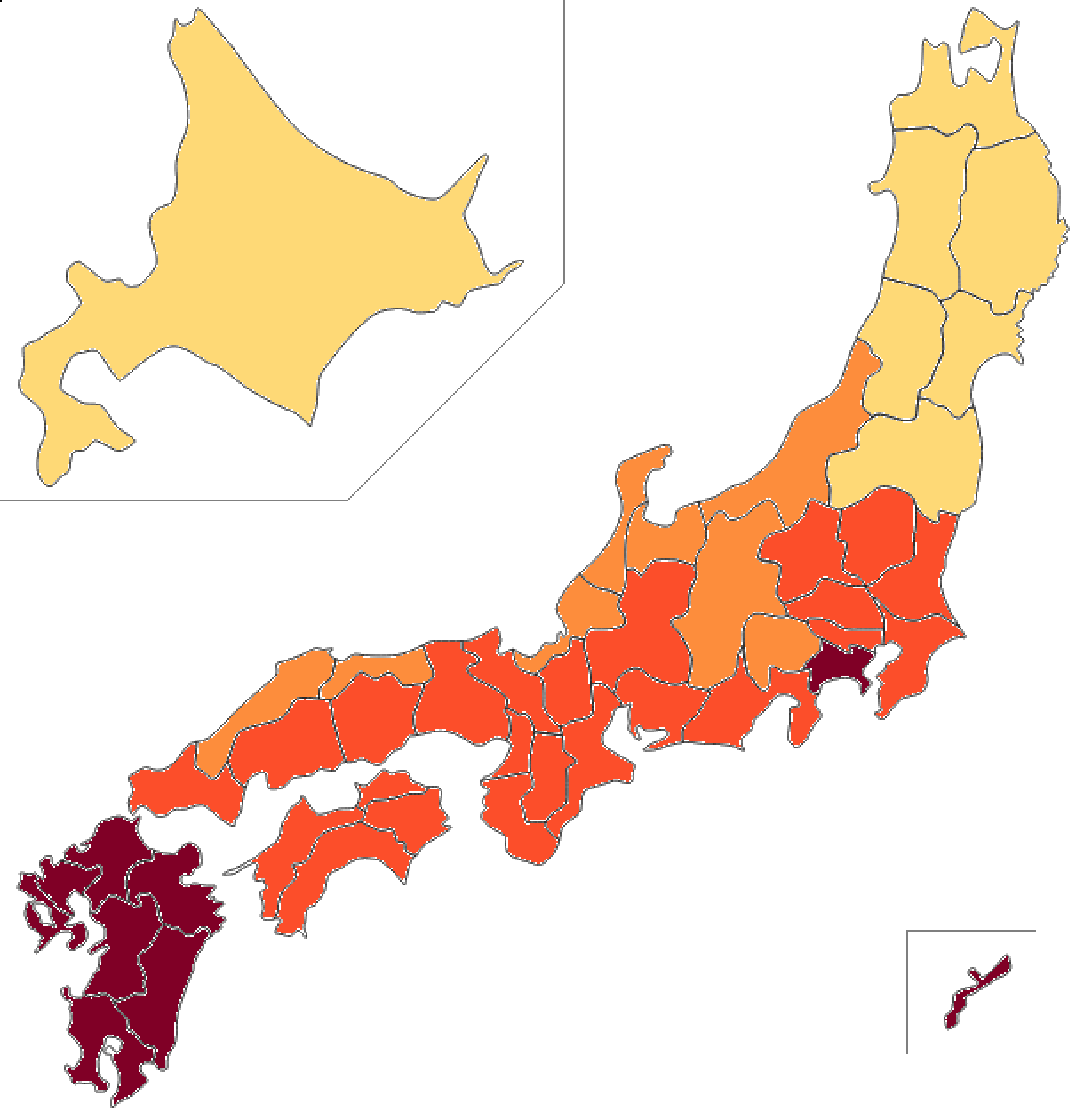
| Region | Climate trends (warming, heavy rain) | Outdoor-sport exposure | Direction of relative risk |
|---|---|---|---|
| Kyushu, Okinawa, Amami | Hot and humid; frequent heavy rain from baiu, typhoons and linear precipitation bands; large rise in sea-surface temperature | High (year-round) | Currently relatively high / clusters already reported |
| Shikoku, Setouchi | Warming; rising probability of "once in 50 years" 3-day precipitation (EA analysis) | High | High → rising |
| Kinki, Tokai, Kii Peninsula (Pacific side) | Hot and humid; more heavy rain; heat-island effect in cities | High | Rising |
| Kanto | More short-duration heavy rain; concentration of large cities; heat island | Very high (large athlete population) | Rising (urban risk emerging); a 2025 cluster reported at a cornea conference |
| Hokuriku, Koshin | More heavy rain; summer warming | Medium–high | Gradually rising |
| Tohoku | Large temperature rise due to high latitude; more heavy rain | Medium | Rising over time (northward shift of suitable areas) |
| Hokkaido | Largest projected temperature rise; more heavy rain; less snow | Medium (mainly summer) | Rising in the long term (previously low-risk) |
II-4. Discussion and Future Challenges
Reviewing the reports and research to date, the positioning of microsporidial keratoconjunctivitis is shifting from a "rare disease" to a "common differential that should be recognized," and in recent years its character as a "climate-sensitive infection," closely linked to climatic and meteorological conditions, has become clear[24, 28]. The remaining challenges are as follows.
- Surveillance: In Japan and elsewhere, a monitoring system and case definitions are needed to grasp the disease's occurrence. Early detection of clusters after outdoor sports is key.
- Environmental surveys: The distribution and persistence conditions of V. corneae in the environment (soil, water, turf) and their relationship to meteorological factors (rainfall, temperature, humidity) need to be clarified.
- Diagnosis and treatment: Standardization of rapid, highly sensitive diagnosis (PCR, mNGS, non-invasive methods) and the establishment of treatment evidence through high-quality randomized controlled trials are required.
- Prevention and adaptation: Preventive education linked to climate-change adaptation is important — avoiding competition on muddy grounds after heavy rain, eye-rinsing and hygiene guidance, eye protection such as goggles, ground drainage and maintenance, and appropriate contact-lens use.
III-1. Analytical Framework — The Meteorological Threshold for Onset and Two Modes of Transmission
1.1 The meteorological threshold
As introduced in Part II (Chapter 2), the study analysing the two Nagasaki outbreaks (16 healthy athletes in total, 2022 and 2023) showed that outbreaks "tend to occur after a month in which the conditions of 23°C temperature, 70% humidity and 200 mm precipitation are met or exceeded."[28] The authors note that heat, high humidity and heavy rain may promote the proliferation of microsporidia in soil and surface water, and state that there is a time lag between the meteorological event and detection of onset, arising from delayed exposure, the incubation period and the stepwise progression of case recognition. A month meeting these three conditions (mean monthly temperature ≥23°C, relative humidity ≥70%, monthly precipitation ≥200 mm) is termed a "high-suitability month" for that region.
Outbreaks tend to occur several months after months meeting these conditions (with a time lag). Real-world risk rises where and when outdoor sports (competition on soil or turf) and water exposure overlap.
1.2 Two modes of transmission (which govern the geographical spread)
Taking the reports as a whole, there are broadly two modes of transmission, and these determine the breadth of the geographical spread.
- The "wet field" type (wet field eye): A route via the soil and surface water of playing fields turned muddy by heavy rain. Typical of tropical-to-subtropical monsoon regions where heat, high humidity and heavy rain coincide, and of temperate-zone summers. The meteorological threshold above applies directly.
- The "water body" type: A route via specific contaminated water bodies such as hot springs, swimming pools, lakes, ponds and rivers (Taiwan's hot springs and pools, Israel's Sea of Galilee, etc.)[12, 13, 14]. Because this type can occur wherever a warm, contaminated water body exists — even if the region's monthly precipitation does not reach the threshold — it extends risk to regions such as Mediterranean climates where "summers are hot but dry."
III-2. Global Climate Change Trends (Key Points from IPCC AR6)
The IPCC Sixth Assessment Report (AR6) states that heavy precipitation increases in frequency and intensity as warming progresses, and that in a 4°C-warmer world, rare heavy-rain events will become more frequent and intense than in the past across all continents and all AR6 regions (virtually certain)[34]. In particular, monsoon precipitation is projected to increase globally over the medium-to-long term, most notably in South and Southeast Asia, East Asia and West Africa[34, 35]. In South Asia, large increases in extreme precipitation are expected in southern India, Myanmar, Thailand, Malaysia and elsewhere[35]. Humid heat (sultry conditions with high wet-bulb temperatures) is also assessed to become stronger and more frequent throughout the 21st century. In short, the heat, humidity and heavy rain that are the conditions for the "wet field" type of this disease are set both to intensify in existing endemic areas and to expand toward their (temperate-side) margins.
III-3. Global Regional / Country-level Outbreak Projections
Combining the fit to the meteorological threshold, opportunities for outdoor-sport and water exposure, and IPCC AR6 future trends, regions and countries are shown in three categories (relative priorities, not probabilities of occurrence).
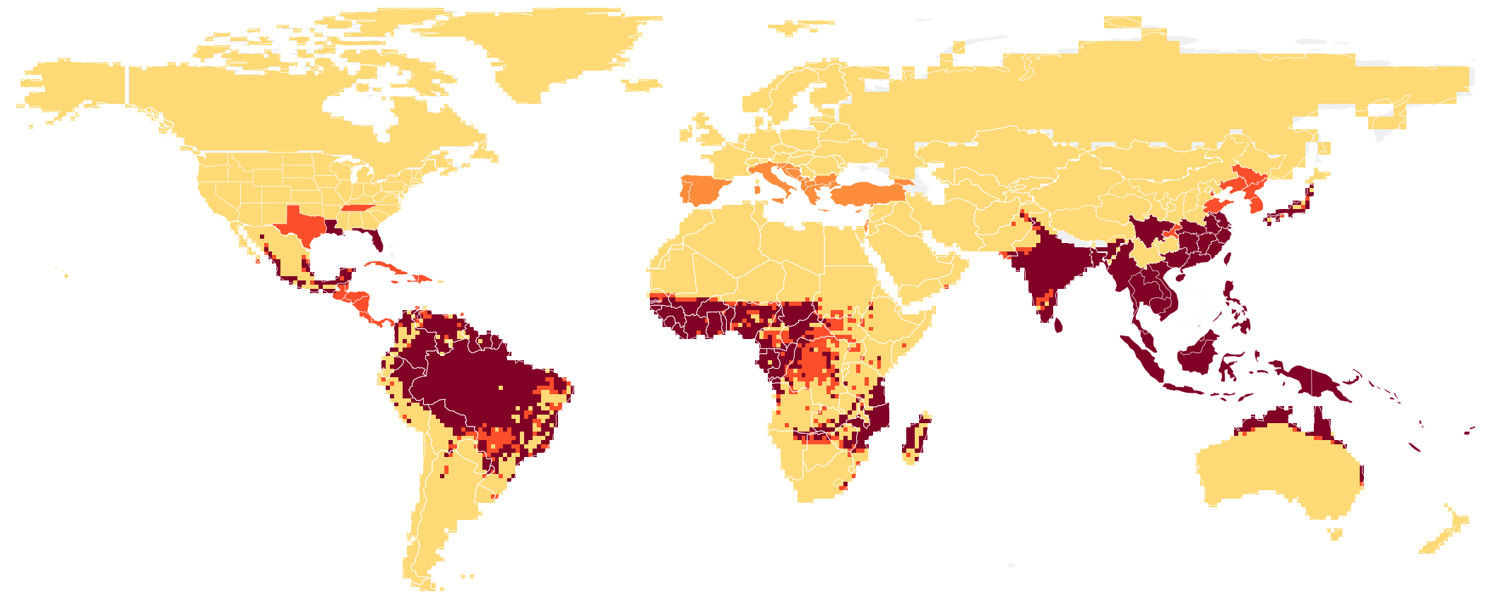
| Category | Region / country | Climate / weather (tendency to meet threshold) | Main exposure / notes |
|---|---|---|---|
| High (endemic / suitable) | Monsoon Asia (South, Southeast, East): India, Bangladesh, Myanmar, Thailand, Vietnam, Malaysia, Indonesia, the Philippines, Sri Lanka, southern China (Guangdong, Hainan, Hong Kong), Taiwan, southern Japan (Kyushu, Okinawa) | Meets the threshold in many months during summer/rainy season. The IPCC projects increased monsoon precipitation here → suitability strengthens further | Mainly the wet-field type. Outdoor sports such as football and rugby are popular. Case series and outbreaks already reported |
| High (endemic / suitable) | Tropical Oceania: northern Australia (Top End / Darwin), Papua New Guinea, Pacific islands | Heat, high humidity and heavy rain in the wet season (November–April in the north). The Darwin outbreak (March) fits the end of the wet season | Outbreak of >100 people in Darwin in March 2026 (V. corneae, Australian-rules football) |
| Expanding (emerging / on alert) | Sub-Saharan Africa monsoon belt: West Africa (Nigeria, Ghana, etc.), Central Africa (Congo Basin, Cameroon), parts of East Africa, coastal Madagascar | Meets the threshold in the wet season (mostly June–September). The IPCC projects increased West African monsoon | Strong football culture, but weak ophthalmic surveillance; likely under-recognized |
| Expanding (emerging / on alert) | Tropical Latin America / Caribbean: Central America (wet season May–October), the Caribbean (hurricane season June–November), Colombia, Venezuela, the Amazon, coastal Brazil (South American monsoon) | Heat, humidity and heavy rain in the wet season. The South American monsoon region has rapidly rising maximum temperatures | Strong football culture. Watch also for the co-existing water-body type (rivers, lakes) |
| Expanding (emerging / on alert) | Temperate East Asian summer: central China (Yangtze basin), South Korea (the "changma" rainy season, June–July), central and northern Honshu (Japan) | Opportunities to meet the threshold increase in summer with warming. In Japan, signs of spread beyond Kyushu (to Honshu, Shikoku, etc.) | Wet-field type. In Japan, cases in golf and rugby in addition to football |
| Local / rising in future | Mediterranean / temperate warm water bodies: Israel (Sea of Galilee); lakes, rivers and reservoirs of southern Europe (Italy, Greece, Spain, Turkey, the Balkans) | Summers are hot but dry, so the precipitation threshold is hard to meet. Mainly the water-body type | Multi-year outbreak in Israel in 2022–2024. Recreational use of warm freshwater is key |
| Local / rising in future | Temperate Europe (central/western Europe, the UK), temperate inland North America (Midwest, Northeast) | Summers warm and short-duration heavy rain increases, but monthly means rarely reach the threshold | Can occur locally at playing fields or warm water bodies just after heavy-rain events. Risk rises with warming |
III-4. Seasonality of Outbreaks (Monthly Risk Calendar)
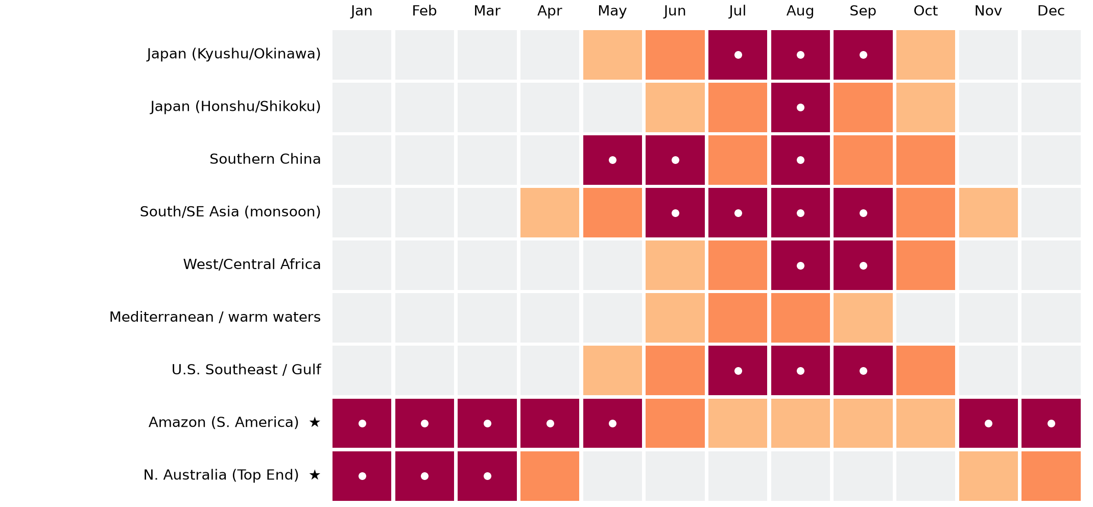
For principal regions, relative outbreak risk is shown by month, based on the meteorological threshold (23°C, 70%, 200 mm)[28] and known seasonality. Northern-Hemisphere regions rise in summer/rainy season (roughly June–September), while northern Australia (★) in the Southern Hemisphere peaks about half a year offset, in January–March. Because there is a lag between meteorological conditions and onset, actual cases may become apparent several weeks after each peak month.
III-5. Positioning the Darwin (Australia) Outbreak
The March 2026 cluster in Darwin is consistent with the framework of this report[31]. The wet season in northern Australia (the Top End) is roughly November–April, and March falls at the end of the wet season — a time when the three conditions of heat, high humidity and heavy rain coincide. Australian-rules football, an outdoor sport on soil and turf, is thought to have generated exposure on the muddied ground. Similar conditions are common to the "high" category above (monsoon Asia, tropical Oceania), and outbreaks of the same kind may recur in these regions. Moreover, the Darwin case shows that the disease also emerges in the Southern-Hemisphere tropics, corroborating the expansion of suitable areas in both north and south directions under climate change.
III-6. Important Caveats for the Projection (Limitations)
III-7. Recommendations (Public Health and Clinical)
- Surveillance: In high-risk regions and periods (especially 4–8 weeks after heavy-rain events), establish systems to detect keratoconjunctivitis clusters in outdoor-sport groups early. Include this disease in the differential for keratoconjunctivitis that is culture-negative and unresponsive to standard treatment.
- Environmental surveys: In both endemic and emerging areas, investigate the presence of V. corneae in playing-field soil and surface water and in recreational warm water bodies (lakes, ponds, hot springs, pools), and its relationship to meteorological factors.
- Prevention and adaptation: In tandem with climate-change adaptation, promote avoiding competition on muddy grounds after heavy rain, eye-rinsing and hygiene after play, eye protection such as goggles, improved ground drainage, appropriate disinfection of pools and hot springs, and appropriate contact-lens use.
- Awareness: Use plain-language terms such as "wet field eye" to raise awareness among players, coaches, the public and clinicians.
III-8. Conclusion
Once regarded as a rare opportunistic infection centred on AIDS patients, microsporidial keratoconjunctivitis has, since the 2000s, been reported with rapidly increasing frequency as a cause of keratoconjunctivitis in immunocompetent individuals, centred on Asia, and its occurrence has been quantitatively shown to be strongly associated with heat and heavy rain. In Japan, two outbreaks were reported in Nagasaki in 2022 and 2023, and the same Vittaforma corneae was detected in patients' corneal specimens and in soil from the playing fields, demonstrating a direct link between the environmental reservoir and human infection[27, 28]. According to JMA data, rising temperatures and increasing frequency and intensity of heavy rain are observed and projected in Japan, and the underlying conditions for this disease are being raised nationwide. By region, risk is relatively high in areas that are hot and humid with frequent heavy rain — Kyushu, Okinawa, Shikoku, Setouchi and Tokai — and, as warming progresses, suitable areas are expected to expand into Kanto, Tohoku and Hokkaido. However, this is a qualitative projection based on limited data, and strengthening of surveillance and environmental surveys is essential. In ophthalmic practice, an attitude of including this disease in the differential is required for keratoconjunctivitis/keratitis that is culture-negative and unresponsive to standard treatment, especially in cases with a history of outdoor sports after heavy rain.
Internationally as well, using the meteorological threshold identified by the Nagasaki study (mean monthly temperature 23°C, relative humidity 70%, monthly precipitation 200 mm)[28] to gauge suitable areas, the monsoon belts of South and Southeast Asia and tropical Oceania are already high-risk, while in the United States the Southeast and Gulf Coast are the top priority. Climate change expands both the regions and the periods that meet this threshold, and is expected to spread outbreaks even into temperate areas previously regarded as "non-endemic." This disease merits cross-disciplinary attention — not only as an ophthalmic condition but as a "climate-sensitive infection" to be monitored under climate change.
III-9. References
- Weber R, Bryan RT, Schwartz DA, et al. Human microsporidial infections. Clin Microbiol Rev. 1994;7:426–461.
- StatPearls. Microsporidial Keratitis. NCBI Bookshelf (NIH), 2024.
- Sharma S, Das S, Joseph J, et al. Microsporidial keratitis: need for increased awareness. Surv Ophthalmol. 2011;56:1–22.
- Ashton N, Wirasinha PA. Encephalitozoonosis (nosematosis) of the cornea. Br J Ophthalmol. 1973;57:669–674.
- Lowder CY, Meisler DM, McMahon JT, et al. Microsporidia infection of the cornea in a man seropositive for HIV. Am J Ophthalmol. 1990;109:242–244.
- Silverstein BE, Cunningham ET Jr, Margolis TP, et al. Microsporidial keratoconjunctivitis in a patient without HIV infection. Am J Ophthalmol. 1997;124:395–396.
- Theng J, Chan C, Ling ML, et al. Microsporidial keratoconjunctivitis in a healthy contact lens wearer without HIV infection. Ophthalmology. 2001;108:976–978.
- Chan CM, Theng JT, Li L, et al. Microsporidial keratoconjunctivitis in healthy individuals: a case series. Ophthalmology. 2003;110:1420–1425.
- Loh RS, Chan CM, Ti SE, et al. Emerging prevalence of microsporidial keratitis in Singapore. Ophthalmology. 2009;116:2348–2353.
- Joseph J, Sridhar MS, Murthy S, et al. Clinical and microbiological profile of microsporidial keratoconjunctivitis in southern India. Ophthalmology. 2006;113:531–537.
- Tan J, Lee P, Lai Y, et al. Microsporidial keratoconjunctivitis after rugby tournament, Singapore. Emerg Infect Dis. 2013;19:1484–1486.
- Fan NW, Wu CC, Chen TL, et al. Microsporidial keratitis in patients with hot springs exposure. J Clin Microbiol. 2012;50:414–418.
- Wang WY, Chu HS, Lin PC, et al. Outbreak of microsporidial keratoconjunctivitis associated with water contamination in swimming pools in Taiwan. Am J Ophthalmol. 2018;194:101–109.
- Friehmann A, Lubitz I, El Zhalka F, et al. Microsporidial keratoconjunctivitis caused by Vittaforma corneae, Sea of Galilee, Israel, 2022–2024. Emerg Infect Dis. 2025;31(8).
- Zhang Y, Xu S, Xu Y. Clinical Analyses of 4 Cases of Microsporidial Keratoconjunctivitis. Acta Parasitol. 2025;70:71.
- Liang R, Zhang B. Bilateral microsporidial keratoconjunctivitis in an orthokeratology lens wearer: a case report and literature review. BMC Ophthalmol. 2026. doi:10.1186/s12886-026-05033-y.
- Das S, Sharma S, Sahu SK, et al. Diagnosis, clinical features and treatment outcome of microsporidial keratoconjunctivitis. Br J Ophthalmol. 2012;96(6):793–795.
- Kodjikian L, Garweg JG, Nguyen M, et al. Intraocular microsporidiosis due to Encephalitozoon cuniculi in a patient with idiopathic CD4+ T-lymphocytopenia. Int J Med Microbiol. 2005;294:529–533.
- Pariyakanok L, Satitpitakul V, Laksanaphuk P, et al. Stromal keratitis with endophthalmitis caused by Vittaforma corneae in an immunocompetent patient: a case report. Ocul Immunol Inflamm. 2019;27(5):826–828.
- Das S, Sahu SK, Sharma S, et al. Clinical trial of 0.02% polyhexamethylene biguanide versus placebo in the treatment of microsporidial keratoconjunctivitis. Am J Ophthalmol. 2010;150:110–115.
- Das S, Wallang BS, Sharma S, et al. The efficacy of corneal debridement in the treatment of microsporidial keratoconjunctivitis: a prospective randomized clinical trial. Am J Ophthalmol. 2014;157:1151–1155.
- Lekskul M, Sasiprapha N, Mungthin M, et al. Clinical characteristics, progression patterns and treatment outcomes in microsporidial keratoconjunctivitis: a prospective study in Thailand. Int Ophthalmol. 2024;44:432.
- Reddy AK, Balne PK, Garg P, et al. Is microsporidial keratitis a seasonal infection in India? Clin Microbiol Infect. 2011;17:1114–1116.
- Das AV, Basu S. Temporal trend of microsporidial keratoconjunctivitis and correlation with environmental and air pollution factors in India. Indian J Ophthalmol. 2021;69:1089–1094.
- Tomooka M, Suzuki T, Toriyama K, et al. A Case of Microsporidial Keratitis Accompanied with Fungal Keratitis. Atarashii Ganka (Journal of the Eye). 2014;31(5):737–741 (in Japanese).
- Ueno S, Eguchi H, Hotta F, et al. Microsporidial keratitis retrospectively diagnosed by ultrastructural study of formalin-fixed paraffin-embedded corneal tissue: a case report. Ann Clin Microbiol Antimicrob. 2019;18(1):17.
- Uematsu M, Mohamed YH, Kusano M, et al. Microsporidial keratoconjunctivitis – first outbreak in Japan. BMC Infect Dis. 2023;23:752.
- Mohamed MT, Uematsu M, Mohamed YH, et al. Environmental and Climatic Drivers of Microsporidial Keratoconjunctivitis in Athletes: Molecular Evidence from Outbreaks in Japan. Microorganisms. 2026;14(3):587. doi:10.3390/microorganisms14030587.
- World Weather Online. Historical weather data (https://www.worldweatheronline.com/). Used to reference the meteorological conditions at outbreak locations and times.
- Kakisu K, Suzuki T, Itokawa T, et al. Cluster infection of Microsporidia keratoconjunctivitis in athletes of a sports team. Japan Cornea Conference 2025 (49th Annual Meeting of the Japan Cornea Society / 41st Annual Meeting of the Keratoplasty Society of Japan), Koyasan University, February 2025. Poster P29 (conference presentation, not yet published).
- Optometry Today (AOP). Expert: climate change may influence frequency and spread of parasitic eye infection. 21 May 2026 (interview with Associate Professor Uematsu; the meteorological threshold of 23°C / 70% / 200 mm and the Darwin outbreak).
- MEXT / Japan Meteorological Agency. Climate Change in Japan 2025 — Assessment Report on Observations and Projections of the Atmosphere, Land and Ocean. 2025.
- Japan Meteorological Agency. Climate Change Monitoring Report (updated 2025/2026); "Annual mean temperature of Japan"; "Past changes in extreme events such as heavy rain and extremely hot days."
- IPCC. Climate Change 2021: The Physical Science Basis (AR6 WGI). Summary for Policymakers (B.2.4 intensification of heavy rain; B.3.3 increase in monsoon precipitation: South and Southeast Asia, East Asia, West Africa); Chapter 11; regional fact sheet (Asia).
- IPCC AR6 WGII Chapter 10 (Asia): frequent and increasing extreme precipitation and flooding in monsoon Asia.
This report is a summary of published academic literature and official data, and its region-level projections are assumption-dependent qualitative outlooks. For diagnosis and treatment and for disaster-prevention and policy decisions, consult the latest primary sources, specialist physicians and the relevant authorities. This document does not constitute medical or administrative advice.
Principal abbreviations
- MKC
- Microsporidial keratoconjunctivitis
- MSK
- Microsporidial stromal keratitis
- V. corneae
- Vittaforma corneae (microsporidian that causes ocular infection)
- IVCM
- In vivo confocal microscopy
- AS-OCT
- Anterior segment optical coherence tomography
- PCR
- Polymerase chain reaction
- mNGS
- Metagenomic next-generation sequencing
- TEM
- Transmission electron microscopy
- PHMB
- Polyhexamethylene biguanide
- PKP
- Penetrating keratoplasty
- ART / HAART
- Antiretroviral therapy
- IPCC AR6
- Intergovernmental Panel on Climate Change, Sixth Assessment Report
- RCP
- Representative Concentration Pathway (emissions scenario)
- EA
- Event attribution (analysis of warming's contribution to extremes)
- JMA
- Japan Meteorological Agency
List of figures and tables
- Table 1. Principal microsporidia involved in ocular infection (Part I, I-2)
- Table 2. Principal diagnostic methods and their features (Part I, I-6)
- Table 3. Region-level outbreak risk in Japan under climate change (Part II, II-3)
- Table 4. Outbreak risk by global region/country (Part III, III-3)
- Figure 1. Region-level outbreak risk in Japan under climate change (Part II, II-3)
- Figure 2. Global outbreak risk (Part III, III-3)
- Figure 3. Monthly outbreak risk for key regions (Part III, III-4)